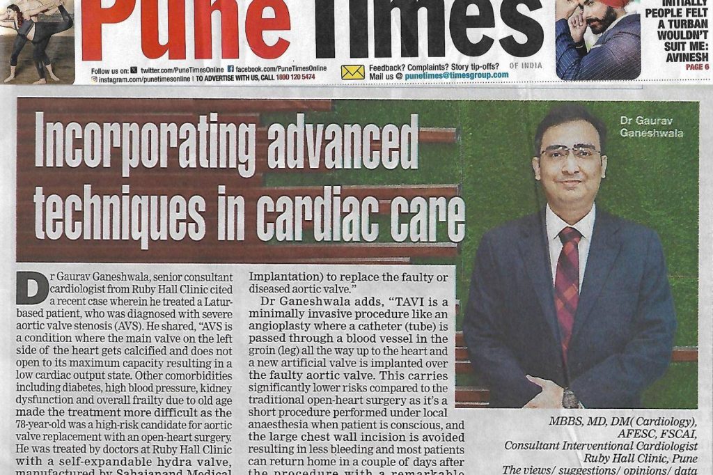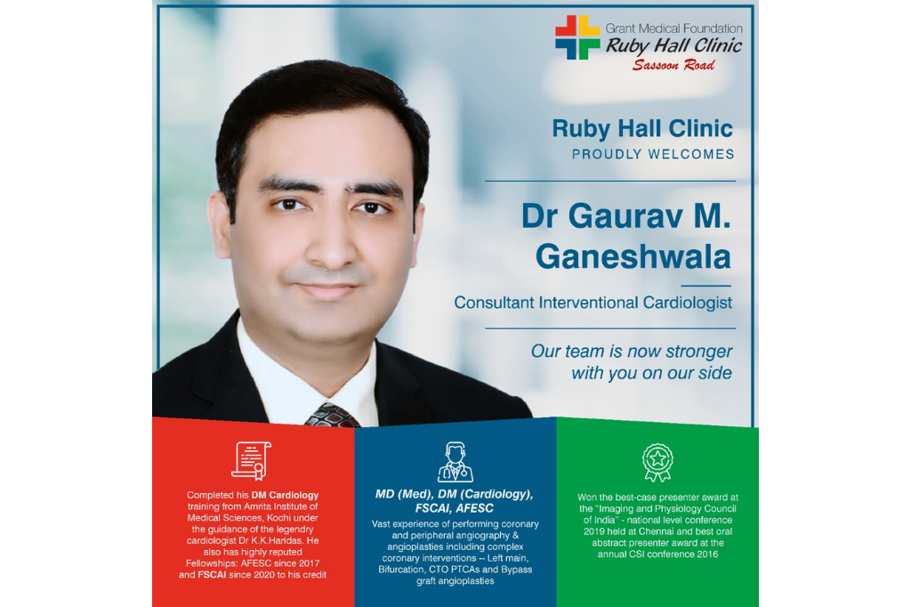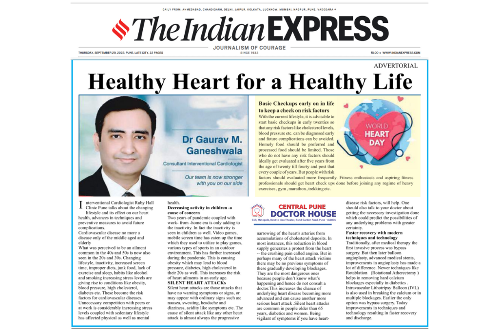
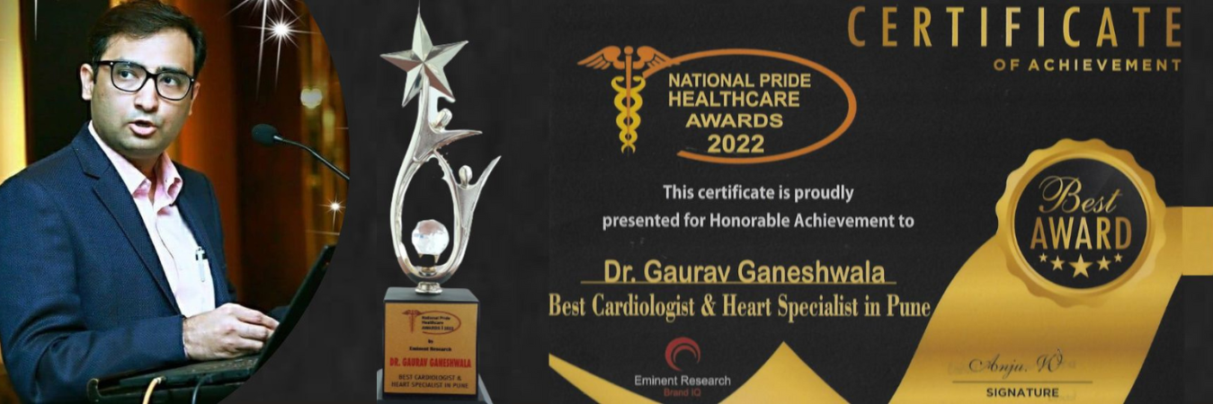
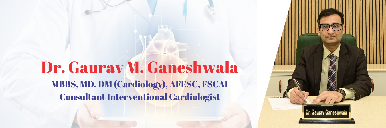


Dr. Gaurav Ganeshwala
MBBS, MD (Medicine), DM (Cardiology), FESC, FSCAI
Senior Consultant Cardiologist & Heart Failure Specialist
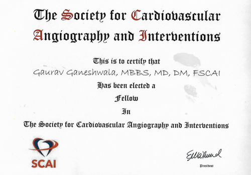
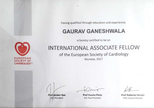
Dr. Gaurav Ganeshwala is a senior Consultant Cardiologist with more than 10 years of experience in the field of clinical and Interventional cardiology. He had completed his DM Cardiology training at the prestigious Amrita Institute of Medical Sciences, Kochi. He also has the honor of International fellowships from the European Society of Cardiology (FESC) and Society for Cardiovascular Angiography and Interventions (FSCAI) to his credit.


Our Services
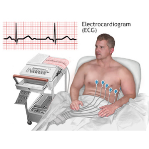
Electrocardiogram (ECG)
An electrocardiogram (ECG) is a simple test that can be used to check your heart’s rhythm and electrical activity.
Echocardiography
(2D ECHO)
An echocardiogram checks how your heart’s chambers and valves are pumping blood through your heart.
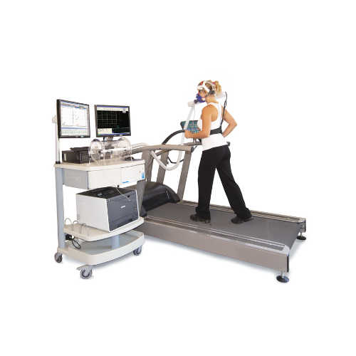
Cardio Pulmonary
Exercise Test (CPET)
Cardiopulmonary Exercise Testing (CPET) is a non-invasive method used to assess the performance of the heart and lungs at rest and during exercise.
Complex Angioplasty
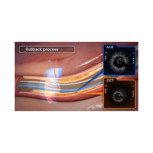
Intracoronary Imaging IVUS, OCT
Intracoronary imaging techniques (intravascular ultrasound (IVUS) and optical coherence tomography (OCT))
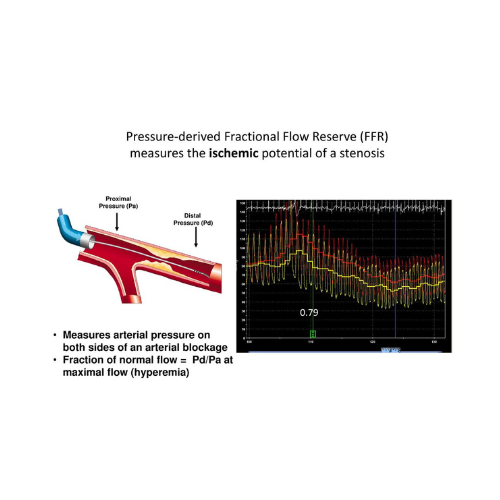
Intracoronary Physiology FFR
Fractional Flow Reserve, or FFR, is a guide wire-based procedure that can accurately measure blood pressure and
Transcatheter Aortic Valve Replacement (TAVR)
Transcatheter aortic valve replacement (TAVR) is a minimally invasive procedure to replace a narrowed aortic valve that fails to open properly.
Transcatheter Mitral Valve Repair (TMVR)
MitraClip
MitraClip is a breakthrough innovation for mitral valve patients. It is a device used to treat mitral valve regurgitation. This minimally invasive, transcatheter approach
Rotablation
Rotablation represents an addition to the standard PTCA procedure and uses a tiny drill, powered by compressed air, to remove calcified deposits.
Bioresorbable Vascular Scaffold
The BVS a non metallic mesh tube that is used to treat a narrowed artery, is similar to a stent, but slowly dissolves once the blocked artery can
Transradial Intervention
The transradial technique is an effective, minimally invasive approach to perform coronary and peripheral angiograms and interventions.
Doctor Consultation
Central Pune Doctor House, Pune
Address: G1B Metropole, Next to Inox Theater, Bund Garden Road, Pune, Maharashtra 411001
Timing :
12 PM to 2 PM (By appointment)
5:30 PM to 7:30 PM
Monday to Saturday
Ruby Hall Clinic, Sasoon Road, Pune
Address: 40, Sassoon Road, Cardiology OPD-2, Ground Floor, Pune, Maharashtra 411001
Timing : 12 PM to 2 PM
Tuesday
Ruby Hall Clinic, Wanowrie, Pune
Address: 59/6, Disney Park, Azad Nagar, Wanowrie, Pune, Maharashtra 411040
Timing :
4 PM to 5 PM
Monday, Tuesday, Thursday and Friday
Make an appointment with
Dr. Gaurav Ganeshwala
Advanced, Evidence-based Heart Treatment
Patient Happy Stories
EXCELLENT
Gallery
Video
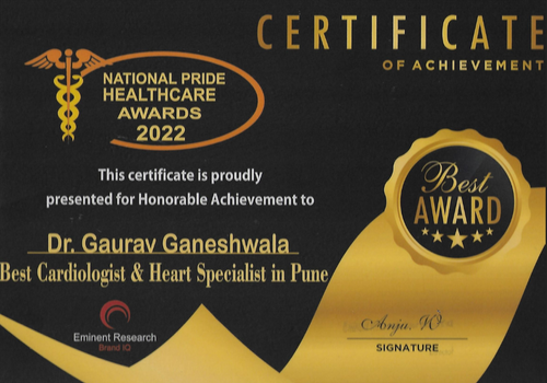
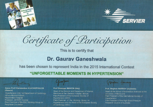
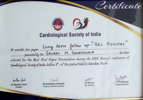
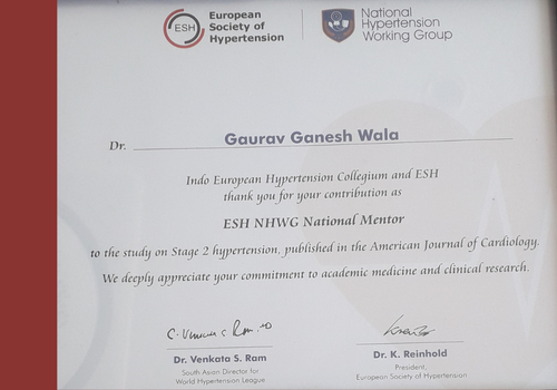
Achievements:
POPULAR SEARCHES
Case of the week
Frequently Asked Questions
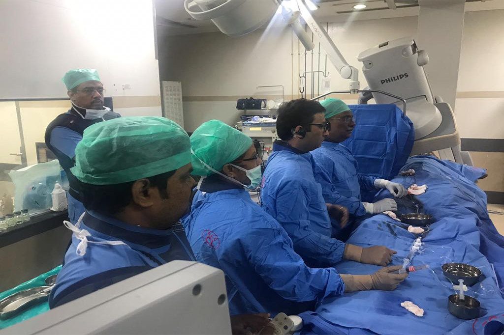
Coronary angioplasty also called PCI or PTCA – is a noninvasive procedure that helps treat coronary heart disease [blocked coronary arteries] by improving the blood supply to the heart muscle, through widening and opening of the narrowed coronary arteries. It is used to stop heart attacks in progress, treat chest pain (angina), and restore blood flow through the coronary arteries. The procedure is performed in the cardiac catheterization laboratory (or cath lab) by a specialized Interventional cardiologist.
An angiogram is performed prior to angioplasty. Here, a thin plastic tube is inserted into an artery in the groin or arm. A long, narrow, hollow tube, called a catheter, is passed through the sheath and guided up the blood vessel to the arteries surrounding the heart. A small amount of contrast liquid is injected through the catheter and is photographed with an X-ray as it moves through the heart’s chambers, valves, and major vessels. From the digital pictures of the contrast material, the doctors can tell whether the coronary arteries are narrowed and whether the heart valves are working correctly.
Intravascular imaging has revolutionized the precision of angioplasty. One type of imaging is the Intravascular Ultrasound, where a miniature probe is used to study the nature of the plaque. Where regular angiography shows only a two-dimensional silhouette of the interior of the coronary arteries, IVUS visualizes the coronary artery from the inside out. This unique point-of-view picture, generated in real-time, yields valuable information.
Yet another innovative method of intravascular imaging is Optical Coherence Tomography [OCT]. This produces high-resolution intracoronary images using infrared light.
The new imaging technologies give crucial information whether the plaque blocking the vessel is hard or soft, is made up of lipids or calcium, etc. They can also give accurate detail about the size of the stent that may be needed, and assess post stenting status of the vessel as well.

