
Intracoronary Imaging IVUS, OCT
Intracoronary Imaging IVUS, OCT
- Intracoronary imaging techniques (intravascular ultrasound (IVUS) and optical coherence tomography (OCT)) are routinely available to complement angiography in the management of coronary artery disease
- Both IVUS and OCT are superior to angiography for quantification of vessel dimension and thus critically helpful in guiding coronaryangioplasty (percutaneous coronary intervention (PCI)) and stent implantation
Intravascular imaging has revolutionized the precision of angioplasty. One type of imaging is the Intravascular Ultrasound, where a Cardiologist uses a miniature probe to study the nature of the plaque. Where regular angiography shows only a two-dimensional silhouette of the interior of the coronary arteries, IVUS visualizes the coronary artery from the inside out. This unique point-of-view picture, generated in real-time, yields valuable information.
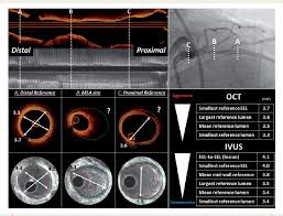
Yet another innovative method of intravascular imaging is Optical Coherence Tomography [OCT]. This produces high-resolution intracoronary images using infrared light. The new imaging technologies give crucial information whether the plaque blocking the vessel is hard or soft, is made up of lipids or calcium, etc. They can also give accurate detail about the size of the stent that may be needed, and assess post stenting status of the vessel as well.
IVUS
Intravascular ultrasound (IVUS) is an imaging methodology using a specially designed catheter with a miniaturized ultrasound probe attached to the distal end of the catheter.
IVUS is performed by inserting a catheter containing a miniaturised probe into the coronary artery {heart blood vessel) which gives highly magnified detailed information of the blood vessel, its wall and helps understand the nature, type and extent of plaques (blocks). This greatly enhances the understanding of the problem and helps to treat the blocks in a safe and effective manner.
IVUS is usually done during percutaneous coronary intervention, such as stenting. IVUS helps to achieve best results after stenting to enhance good long term outcomes.
During complex stenting procedures like left main, bifurcation and chronically occluded vessels. It is also useful to assess failed stents.
Dr Gaurav Ganeshwala, Ruby Hall Clinic, Pune is one of the pioneers and highly experienced IVUS operator in the country.
OCT
Optical coherence tomography (OCT) is an intracoronary imaging technique that produces high resolution images of the blood vessel and its walls. Optical coherence tomography is used to assess vessel size, the nature, type, distribution of blocks. It is nowadays used more as a guide during coronary stenting procedures. OCT helps to achieve stenting results with precision as the lumen is visualised in a magnified view similar to electron microscopy.
OCT uses infrared light to image the blood vessels and the entire blood vessel can be imaged in 3 second scan. The wavelength of light is much shorter and much faster than sound waves and hence OCT images have a resolution 10 times greater than IVUS. The catheter with infrared emitting tip is advanced into the blood vessel and images are acquired while the dye is injected.
Optical coherence tomography is used to obtain detailed evaluation of coronary atherosclerotic plaques and the vascular response to coronary interventional devices, such as stents. Optical coherence tomography can be used as a guide during coronary intervention.
Dr Gaurav Ganeshwala is key opinion leader of OCT in India and is highly experienced in OCT.